
Abstract and Introduction
Abstract
Osteoporosis is a disease characterized by low bone mass and microarchitectural deterioration leading to increased bone fragility and fracture risk. Typically, osteoporotic fractures occur at the spine, hip, distal forearm, and proximal humerus, but other skeletal sites may be affected as well. One of the major challenges in the management of osteoporosis lies in the fact that although the operational diagnosis is based on bone mineral density (BMD) as measured by dual x-ray absorptiometry, the majority of fractures occur at nonosteoporotic BMD values. Furthermore, osteoporosis often remains undiagnosed regardless of the low severity of the underlying trauma. Also, there is only weak consensus among the major guidelines worldwide, when to treat, whom to treat, and which drug to use. Against this background, increasing efforts have been undertaken in the past few years by artificial intelligence (AI) developers to support and improve the management of this disease. The performance of many of these newly developed AI algorithms have been shown to be at least comparable to that of physician experts, or even superior. However, even if study results appear promising at a first glance, they should always be interpreted with caution. Use of inadequate reference standards or selection of variables that are of little or no value in clinical practice are limitations not infrequently found. Consequently, there is a clear need for high-quality clinical research in this field of AI. This could, eg, be achieved by establishing an internationally consented "best practice framework" that considers all relevant stakeholders.
Introduction
Osteoporosis is defined as a systemic skeletal disease characterized by low bone mass and microarchitectural deterioration of bone tissue with a consequent increase in bone fragility and susceptibility to fracture.[1] According to criteria recommended by the World Health Organization (WHO), the operational diagnosis of osteoporosis is based on bone mineral density (BMD) as assessed by dual x-ray absorptiometry (DXA) of the hip, the spine, or the so-called one-third radius.[2,3] Osteoporosis can thus be diagnosed if an individual's BMD is equal to or less than −2.5 standard deviations below the mean normal BMD value of healthy young adults (ie, at a T-score ≤ −2.5). However, one of the main limitations of this approach lies in the fact that the majority of fractures occur at T-scores of −1.0 to −2.5 (ie, at osteopenic BMD) or even above −1.0 (ie, at normal BMD), compromising the sensitivity of this "gold-standard" method and its potential role as a screening tool.[4] Fractures at the spine (vertebrae), hip (proximal femur), shoulder (proximal humerus), and wrist (distal forearm, distal radius) have been shown to be associated with an increased risk of subsequent fracture, reduced quality of life, disability, and, except for distal forearm fracture, increased mortality.[5–8] Accordingly, they have also been referred to as major osteoporotic fractures. The average lifetime risk of a 50-year-old woman to suffer a major osteoporotic fracture has been estimated at close to 50%, and in men 22%, and, worldwide, osteoporosis causes some 9 million fractures annually, resulting in an osteoporosis fracture every 3 seconds.[9,10]
The term artificial intelligence (AI) in its current sense was most likely coined in the mid-1950s, when a group of mathematicians and cognitive and computer scientists convened at a conference at Dartmouth College (U.S.). While the conference itself fell short of the participants' expectations, it can nevertheless be considered the initial spark for the AI research boom that followed. This boom, however, was interrupted at least twice by periods of research slump, sometimes also referred to as "winters of AI research," the first of which lasted from the mid- to the late 1970s and the second from the late 1980s to the early 1990s. Both "winters" were preceded by barely encouraging research results, which in turn led to a decrease in funding of AI-related research projects. Fortunately, with an almost exponential growth of computational power, research and funding began to pick up again after that. In 1997, an IBM computer called IBM Deep Blue® beat the world chess champion Gary Kasparov, and in 2011 another IBM computer, called Watson®, beat 2 of the all-time most successful human players of the game Jeopardy in front of millions of television viewers. Undoubtedly, these and many subsequent highlights in the development of AI have formed the perfect foundation for AI-based research efforts in human medicine. In fact, substantial progress across many areas of human medicine has been made in the past decade.[11,12] In general, AI in medicine can be divided into 2 subtypes, namely virtual and physical, with the former including eg, imaging solutions and treatment decision support tools, and the latter, eg, intelligent prosthesis and robot-assisted surgery.[13] In regard to the management of osteoporosis, the virtual subtype of AI currently plays the main role, with solutions available (or in development) to facilitate diagnosis, fracture risk assessment, fracture detection, bone quality assessment, and treatment decision[14] (Figure 1).
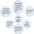
Figure 1.
Selection of currently available artificial intelligence solutions for osteoporosis management.
J Clin Endocrinol Metab. 2023;108(4):775-783. © 2023 Endocrine Society


